
Complete Cure of a Ruptured Cerebellar AVM by Endovascular Embolisation
Introduction
Treatment of symptomatic brain arteriovenous malformations is multimodal, including microsurgical excision, stereotactic radiosurgery, and endovascular embolisation. More often than not, it requires more than one method of treatment.
Case Description
A 38 year old gentleman presented with giddiness, diplopia, and deviation of angle of mouth. On examination, there was a right LMN type of facial palsy and right abducens nerve palsy. There was ataxia in the form of dysdiadochokinesia. No motor weakness noted.
CT and MRI brain revealed bleed in the right middle cerebellar peduncle and adjacent right cerebellar hemisphere. A digital subtraction angiogram was done. The vertebral angiogram revealed AVM nidus with arterial feeders from cerebellar branches of the right anterior inferior cerebellar artery. The AVM nidus measured 2.8 cm x 2.5 x 2.3 cm in size. The venous drainage was into the posterior perimesencephalic veins. There was focal stenosis of the draining vein.

Must Read: Acute stroke treatment at 8 hrs by Interventional Radiologist Dr Suresh Giragani
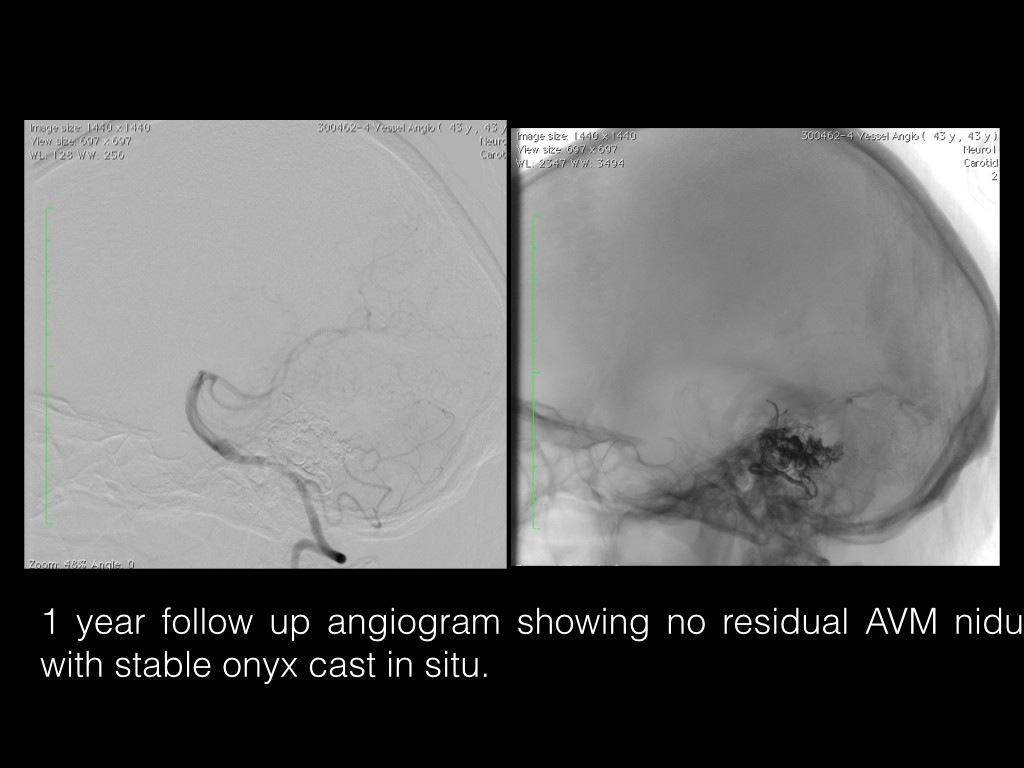
As the AVM nidus was having a single arterial feeder with discrete nidus endovascular embolisation was considered. The embolisation was performed on the seventh day after the ictus. Via right transferral route, the left vertebral artery was accessed and the right AICA feeder selectively cannulated with Apollo detachable tip micro catheter. Onyx-18 embolic agent was used to occlude the fistula. Check angio showed complete occlusion of the AVM nidus. Post procedure, the patient had mild worsening of the facial palsy and ataxia, which was managed by antiplatelets, antiedema measures, and physiotherapy. No increase in the size of hemorrhage noted in the cerebellum. The patient was discharged on 7th post operative day in a stable condition. A cerebral angiogram after 1 year showed no residual AVM nidus. At 1 year follow up patient had mild right sided facial palsy, with no motor weakness.