Pulmonary Artery Embolization Procedurein Hyderabad
What are the diseases treated by bronchial artery and pulmonary artery interventions ?

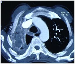
Massive hemoptysis ( coughing large amounts of blood) is a life threatening condition. It is not uncommon and in India, it is commonly caused by long standing lung tuberculosis. Fungal infections in the lung, rarely lung tumors can cause massive hemoptysis.
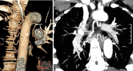
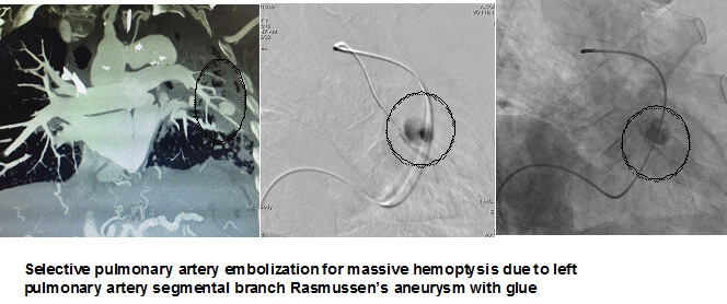
Massive hemotysis is an emergency, may require bronchial artery embolisation if the source of bleeding is from bronchial circulation. All cases of massive hemotysis will undergo CT angiography to see for abnormalities in bronchial and pulmonary circulation.
Pulmonary AVMs are rare pulmonary vascular malformations seen in the lungs. They have abnormal connections between the arteries and veins supplying the lungs. Patients with pulmonary AVM can develop hemoptysis, infections in the brain etc. If detected and symptomatic pulmonary AVMs can be treated by endovascular coil embolisation.


What are the outcomes of bronchial artery embolisation in treating massive hemoptysis ?
Bronchial artery embolization may be life saving in 80-90 % of patients with massive hemoptysis. However, its effect is palliative for symptom relief, the primary disease ( lung infection, lung cavity) needs to be treated by either by surgery or medication.
What is Carotid Cavernous Fistula and Why Does it Matter?
How bronchial artery embolisation is done ?
It is done by inserting small catheters (plastic tubes) in the blood vessel in the groin through a small pinhole incision. These catheters will be taken till the target blood vessel and the embolizing agent ( drug which can close the bleeding vessel) is injected into the bleeding blood vessel in the lung. Damage to the normal lung circulation is avoided during the procedure. The patient needs 6 hours of bed rest after the procedure. He can walk, do routine activities later.
World Stroke Day 2022: Minutes Can Save Lives
What are the emergency bleeding conditions ?
Diseases of various organs can causing bleeding either by invading or involving the blood vessels themselves. Most of the times these episodes of bleeding are manageable by medication or local measures. However, some times if it is torrential, it may be life threatening and it may require an open surgery to close the blood vessel for controlling the bleeding. Embolization technique achieves the same purpose without an open surgery, by selectively closing the bleeding blood vessels through small tubes inserted through the thigh
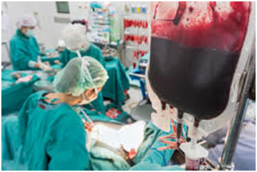
Few examples of emergency bleeding conditions and clinical features
| Symptoms | Site of bleeding | Source |
|---|---|---|
| Massive hemoptysis | Lungs | Bronchial/ pulmonary arteries |
| Postpartum Hemorrhage | Uterus | Uterine arteries |
| Epistaxis | Nose and nasopahrynx | External carotid branches/ internal carotid artery |
| Hematemesis | Upper gastrointestinal tract | Venous and arterial causes |
| Per rectal bleeding | Gastrointestinal tract | Arterial and venous causes |
| Hematuria | Kidney, rarely bladder | Renal arteries |
| Carotid blow out | Oral cavity, oropharynx | Oral cavity, oropharynx External carotid branches/ internal carotid artery |
How do an interventional radiologist treats an acute bleeding condition ?
Many of the emergency bleeding conditions can be treated by interventional radiology in a minimal invasive way. By passing small tubes and selectively cannulating the bleeding vessels, the interventional radiologist will be able to put closing materials (embolic agents) into the involved blood vessels, to close off the vessels. It is relatively safe, noninvasive does not require general anaesthesia and most importantly relatively quickly can be done in an unstable patient.
All You Need To Know About Genicular Artery Embolization For Knee Pain
What are the types of embolizations those can be done in acute bleeding conditions ?
| Symptoms | Type of embolisation |
|---|---|
| Massive hemoptysis | Bronchial artery embolization |
| Postpartum Hemorrhage | Uterine artery embolization |
| Epistaxis | Internal maxillary embolization |
| Hematemesis | Selective arterial embolization for arterial bleeds TIPSS and BRTVO in variceal bleeds |
| Per rectal bleeding | Selective arterial embolization for arterial bleeds |
| Hematuria | Selective renal artery embolization |
| Carotid blow out | Exteranal carotid branch embolisation |
Advantages of Prostatic Artery Embolization for an Enlarged Prostate
Case Example52 year old gentleman had history of oral cavity cancer for which he already underwent radiotherapy & hemimandibulectomy with flap repair, presented to ER with massive oral cavity bleeding due to carotid blow out. He was treated by emergency embolisation of the facial artery and patient was discharged on the 2nd day in a stable condition


Dural arteriovenous fistula (DAVF) Symptoms & Treatment
DR.SURESH GIRAGANI CONSULTANT INTERVENTIONAL RADIOLOGIST at Apollo Hospital, has more than Seventeen years of clinical experience in vascular interventions with a special interest in neurovascular and peripheral vascular disease interventional procedures.
Services
Quick Links
Contact Us
Copyright 2021 Neuro All rights reserved. | Powered By KBK Business Solutions


