
The Ultimate Guide to Aneurysm Coiling

What is a brain aneurysm?
A brain aneurysm is a balloon-like swelling that results from a weakness in the wall of one of the blood vessels supplying blood to the brain. There is a risk that the aneurysm will rupture (burst suddenly) and cause a hemorrhage (bleed).
What is aneurysm coiling?
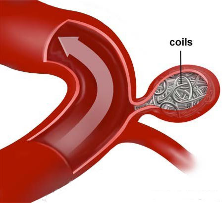
The brain aneurysm is a focal weak part of a brain blood vessel wall, it may sometimes burst and cause bleeding within the brain. The principle of brain aneurysm coiling is to close the aneurysm selectively and preserve the harboring parent vessel blood supply to the brain. This involves fine and meticulous placement of coils within the aneurysm to completely stop the blood flow into the aneurysm so that it is segregated/ excluded from the brain circulation. These coils are delivered through a long thin tube called a microcatheter inserted via the groin.
While viewing an x-ray monitor, called a fluoroscope, the doctor steers the microcatheter through the blood vessels. A special dye injected into the bloodstream makes the blood vessels visible on the monitor. The result is a kind of roadmap for the arteries to be able to be navigated.
When the catheter arrives at the aneurysm, an exceptionally thin and soft platinum coil is delivered. The wire coils up as it enters the aneurysm and is then detached. Different coils are stuffed inside the aneurysm to hinder the bloodstream from entering the aneurysm. After some time, clot forms inside the aneurysm, adequately eliminating the danger of aneurysm rupture. Coils stay inside the aneurysm forever. Coils are made of platinum and they are inert and compatible with the human body.
Aneurysms are variable in size and shape. Saccular aneurysms have a definable neck at their base. Few saccular aneurysms such as wide-necked aneurysms and fusiform/ dissecting aneurysms, don’t have a typical narrow neck. Putting coils into these aneurysms might be complicated and need extra help from stents or balloons. Such kinds of aneurysms is better suited for endovascular flow diverter placement.
Preparation for the procedure of aneurysm coiling
The medical imaging department will give you instructions on preparing for your procedure.
- You may be given blood-thinning medications to take for a few days before your appointment.
- Please tell the staff if you are or suspect you might be pregnant or are breast-feeding
During the procedure
The room will have several super-advanced scanning equipment which is expected to perform the aneurysm coiling.
A medical caretaker will shave a little space of your groin where the catheter will be embedded.
The Interventional neuroradiologist will make a small nick in your groin through which they will introduce the catheter into the artery in your leg. The catheter is then directed through other arteries in your body until it arrives at your brain and then the aneurysm.
The coils are then placed through the catheter, they are sequentially embedded into the aneurysm. The coils are made of platinum, are double the width of a human hair, and can differ long. The quantity of coils required relies upon the size of the aneurysm. The biggest coils are embedded first and afterward, more modest curls are embedded until the aneurysm is filled.
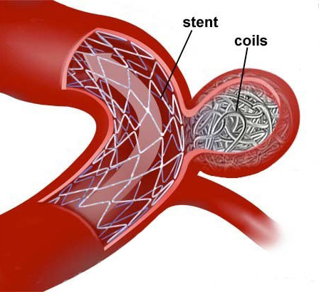
Sometimes, an expandable lattice tube, called a stent, may likewise be placed into the brain blood vessel beneath the aneurysm to hold the coils set up inside the aneurysm. This is called stent-assisted coiling. It is bound to be done assuming the aneurysm has an exceptionally wide ‘neck’ that could some way or another permit the coils to protrude into the parent artery.
When the coiling is complete, the catheter is removed from circulation and the small cut in your groin might be shut by manual compression or by utilizing a tiny ‘plug’ called a vascular closure device. Coiling is a complex and delicate procedure that will take at least three hours and often longer.
After the procedure
Firstly you will be monitored in the Intensive Care Unit (ICU) or High Dependency Unit (HDU), then transferred to the ward. You will need to lie flat and keep your leg (or arm) still and straight for 4 to 6 hours. Moving too soon after the procedure may cause bleeding at the puncture site.
You are encouraged to resume normal daily activities as soon as possible. Staff will discuss with you the level of activity recommended after your coiling.
You will be required to have follow-up imaging procedures (angiograms, MRI, and/or CT) to monitor your aneurysm over the next few years.
What are the risks of coiling?
The risks and complications with this coiling procedure are rare, accounting for less than 5 percent of cases can include but are not limited to the following.
- Minor Pain, and swelling in the groin region.
- Bleeding or swelling might happen in the groin. This is generally halted by applying pressure. This is more common if you’re already taking blood thinners such as Aspirin, Warfarin and Clopidogrel.
- Rarely the patient may have minor or major stroke due to blockage or bleeding of the harboring blood vessel.