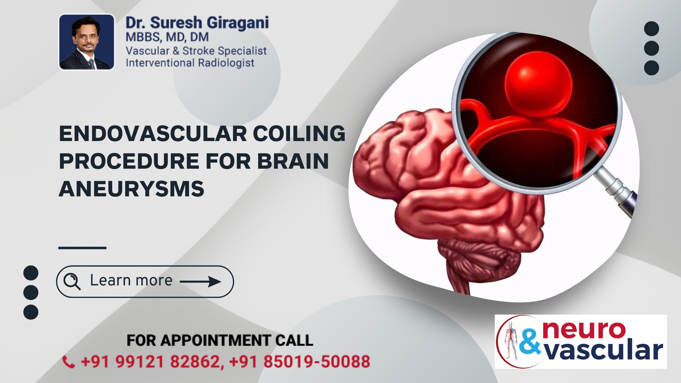
Endovascular Coiling Procedure for Brain Aneurysms

Endovascular coiling is a minimally invasive procedure used to treat cerebral aneurysms, including those located on the anterior communicating artery (ACOM). This procedure aims to prevent an aneurysm from rupturing by sealing it off from the bloodstream.
What is Brain Aneurysm?
The Brain aneurysm occurs when there is a weakened area in the wall of the anterior communicating artery, which connects the left and right anterior cerebral arteries. This weakened area can bulge out and form an aneurysm, which can potentially lead to serious health issues if it ruptures.
What is Endovascular Coiling?
Endovascular coiling is a technique where a catheter is inserted through a small incision, typically in the groin, and navigated to the aneurysm site using imaging guidance. Once in place, small coils are deployed into the aneurysm to induce clotting and prevent blood flow into the aneurysm, effectively reducing the risk of rupture.
Indications for Endovascular Coiling
- Unruptured Brain Aneurysms: To prevent potential rupture and associated complications.
- Ruptured Aneurysms: To control bleeding and stabilize the patient.
Procedure Overview
- Preparation:
- Pre-Procedure Imaging: CT or MRI scans are performed to map the aneurysm and plan the procedure.
- Anesthesia: The patient is typically given local anesthesia with sedation. In some cases, general anesthesia may be used.
- Catheter Insertion:
- Access Site: A small incision is made in the groin, and a catheter is inserted into the femoral artery.
- Catheter Navigation: Using fluoroscopy (real-time X-ray imaging), the catheter is carefully guided through the blood vessels to the site of the aneurysm.
- Coil Deployment:
- Microcatheter Insertion: A smaller catheter is threaded through the initial catheter and directed into the aneurysm.
- Coil Placement: Soft platinum coils are introduced into the aneurysm through the microcatheter. The coils are designed to fill the aneurysm and promote clotting. They can be tightly packed to effectively seal off the aneurysm.
- Completion:
- Final Imaging: Post-procedure imaging, such as angiography, is used to ensure that the aneurysm is adequately occluded and that blood flow in the surrounding vessels is normal.
- Catheter Removal: The catheters are carefully removed, and the incision site is closed.
Recovery and Aftercare
- Hospital Stay: Most patients can go home the next day or after a short hospital stay, depending on their overall condition and any potential complications.
- Follow-Up Imaging: Regular follow-up imaging (such as MRI or angiography) is required to monitor the aneurysm and ensure that it remains occluded.
- Activity Restrictions: Patients are typically advised to avoid strenuous activities and heavy lifting for a period to allow the site to heal properly.
Benefits of Endovascular Coiling
- Minimally Invasive: The procedure requires only a small incision and has a shorter recovery time compared to open surgery.
- Reduced Risk: Effective in reducing the risk of aneurysm rupture and related complications.
- Quick Recovery: Most patients experience a faster recovery and can resume normal activities sooner.
Conclusion
Endovascular coiling is a highly effective treatment for Brain aneurysms, offering a minimally invasive approach to preventing rupture and managing aneurysm-related risks. If you or a loved one has been diagnosed with Brain aneurysm, discuss with a specialist to determine if endovascular coiling is the appropriate treatment option. Regular follow-up and adherence to medical advice are crucial for ensuring the best possible outcomes.
About the Author:

Name: DR . SURESH GIRAGANI
INTERVENTIONAL RADIOLOGIST
DR. SURESH GIRAGANI CONSULTANT INTERVENTIONAL RADIOLOGIST at Apollo hospitals Jubilee Hills has more than sixteen years of clinical experience in vascular interventions with a special interest in neurovascular and peripheral vascular disease interventional procedures.