
A versatile imaging technique applied across five major neurovascular interventions — from diagnosis to the most complex embolization.
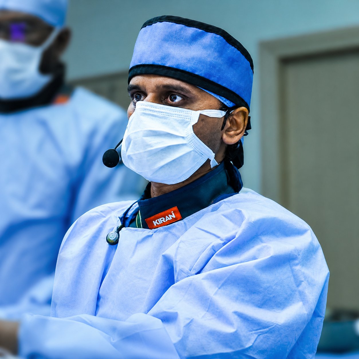
Bi-plane angiography is applied throughout neurovascular intervention for better understanding and execution: cerebral angiography/DSA, stroke thrombectomy, aneurysm coiling and flow diverter placement, AVM embolization, and carotico-cavernous fistula embolization.
Cerebral angiography diagnoses problems within the brain's blood vessels by injecting contrast dye. Though invasive, it's a safe, effective diagnostic tool for detecting aneurysms, arteriovenous malformations, blood clots, and vessel blockages.
Bi-plane angiography completes the procedure in a shorter duration and with a lower contrast dose.
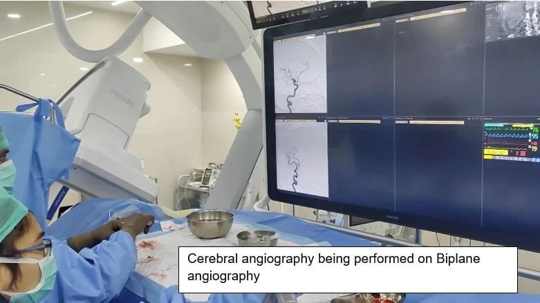
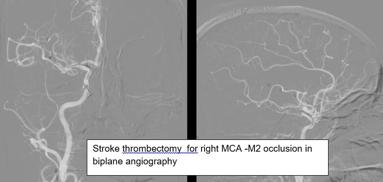
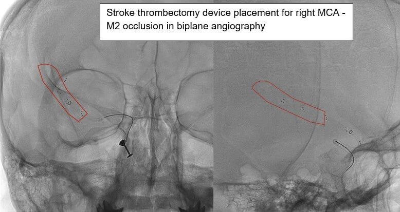
Blockage site and thrombectomy device navigation seen better in two planes at once.
Deeper, smaller vessel blockages cleared more effectively and safely.
Dissections and chronic long-standing occlusions accessed more accurately.
Faster treatment translates directly into better stroke outcomes.
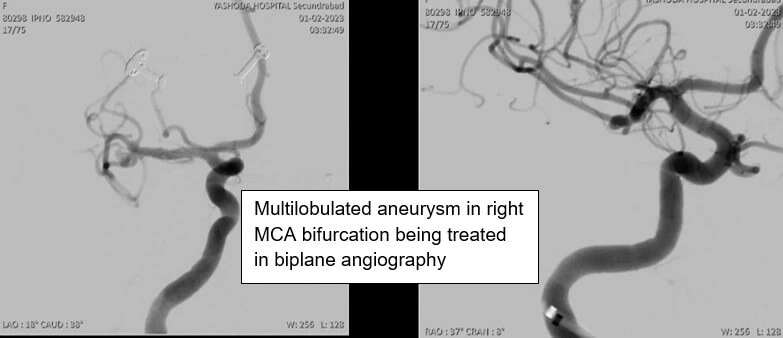
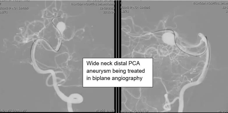
Aneurysm details are exquisitely delineated by simultaneous projections from two fluoroscopic C-arms. This means the entire endovascular procedure is done with utmost accuracy and minimum complication risk.
This is especially valuable for multilobulated aneurysms, complex wide-neck aneurysms, and fusiform aneurysms — whether treated by endovascular coiling or flow diverter placement.
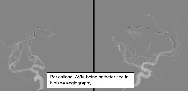
Arteriovenous malformations are abnormal connections between arteries and veins that can lead to haemorrhage, seizures, and neurological deficits. Visualising the AVM's structure and blood flow helps determine the best treatment course.
Dynamic, real-time information about the extent and location of glue percolation within the AVM is essential for safe, effective embolization — provided by simultaneous two-plane imaging.
A carotico-cavernous fistula (CCF) is an abnormal communication between the carotid artery and the cavernous sinus, from trauma or occurring spontaneously. Understanding this complex communication is essential for treatment planning, and endovascular embolization is the only definitive cure.
In some cases, endovascular access requires drilling into adjacent venous sinuses like the inferior petrosal sinus. This complex step can be done safely with bi-plane imaging by mirroring the normal opposite side onto the affected sinus.
Its main applications are cerebral angiography (DSA), stroke thrombectomy, aneurysm coiling and flow diverter placement, brain AVM embolization, and carotico-cavernous fistula embolization.
It allows faster visualisation of the blockage site and more accurate navigation of the thrombectomy device, including for tough-to-clear blockages like dissections and chronic occlusions in deeper, smaller vessels — where speed and accuracy both directly affect outcomes.
Multilobulated, wide-neck, and fusiform aneurysms are exquisitely delineated by two simultaneous fluoroscopic projections, allowing the entire endovascular procedure to be done with maximum accuracy and minimum complication risk.
AVM embolization is considered the most technically challenging neuroendovascular procedure. It requires dynamic, real-time visualisation of exactly where liquid embolic glue is percolating within the AVM, which is only possible with simultaneous two-plane imaging.
A CCF is an abnormal connection between the carotid artery and the cavernous sinus, caused by trauma or occurring spontaneously. Endovascular embolization is the only definitive cure, and bi-plane imaging helps pinpoint the exact location and severity of the fistula for safe treatment.
Dr. Suresh Giragani, Consultant Interventional Radiologist at Apollo Hospitals, Jubilee Hills, Hyderabad, performs all of these procedures using bi-plane angiography, with over 18 years of experience.
Book a consultation with Dr. Giragani, or send your scans and reports for a specialist second opinion.