
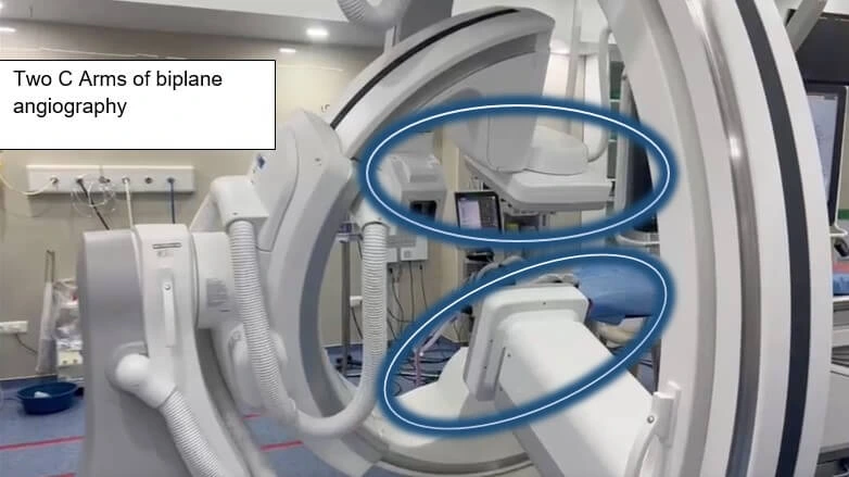
The development of advanced software based on Flat panel X ray for Biplane angiography is a significant step in planning and monitoring of the endovascular treatment within the angiosuite.
This innovative system allows Neurointerventional radiologists to obtain side-by-side and overlapping images of blood vessels and organs.
With detailed 3D and 2D images that can be viewed in real-time, doctors can make quicker and more precise decisions about patient care.
Some of the important techniques are:

3DRA is used to create 3D images of blood vessels in the body. The computed tomography type uses X-rays and a contrast agent to create a detailed, three-dimensional image of the blood vessels.
During a 3DRA procedure, a catheter is inserted into a blood vessel and navigated to the area of interest. A contrast agent is injected through the catheter to fill the blood vessels and make them visible on the X-ray images. The X-ray machine is then rotated around the patient, capturing multiple images from different angles. These images are then processed using specialized software to create a 3D image of the blood vessels.
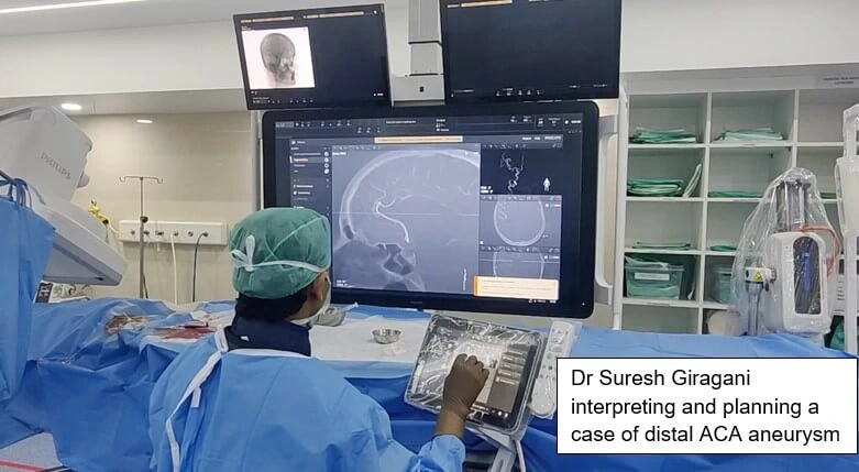
3DRA is commonly used in neuro-interventional procedures to diagnose and treat cerebral aneurysms, arteriovenous malformations, and carotid-cavernous fistulas. It allows interventional neuroradiologists to precisely visualize the anatomy of the blood vessels and plan the most appropriate treatment.
One advantage of 3DRA is that it provides a more detailed and accurate view of the blood vessels compared to traditional 2D angiography. This can be particularly helpful in identifying small aneurysms or malformations that may be difficult to detect with other imaging techniques.
In addition 3D RA helps in planning and execution of endovascular treatment by providing best possible working angles while doing the fluoroscopy.
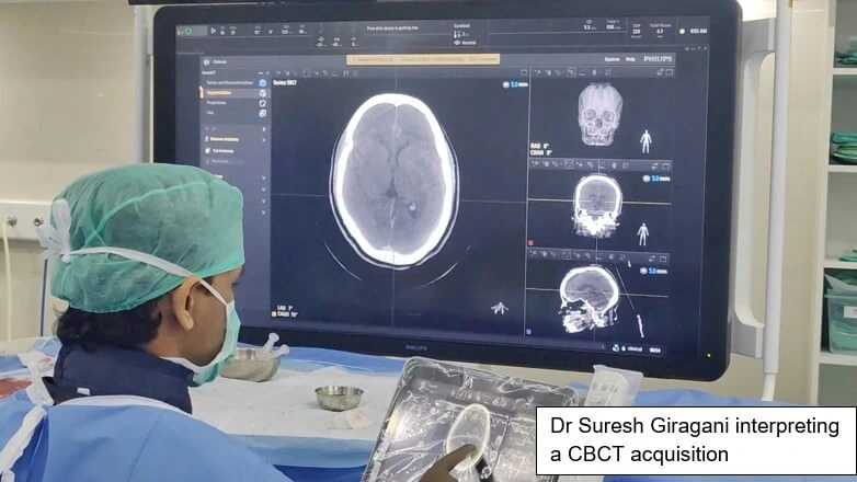
CBCT is used in conjunction with biplane angiography to provide additional information. Also, this helps enhance the accuracy of the 3D reconstruction. By combining the two imaging techniques, interventional neuroradiologists can obtain a more detailed view of the vasculature and surrounding structures. The images are processed using specialized software to create a 3D vasculature reconstruction during a biplane angiography
CBCT can complement biplane angiography by providing additional information about the surrounding structures, such as bones and soft tissues, those may not be visible on the angiography images. This can help interventional neuroradiologists to understand the anatomy better and plan their procedures more accurately
CBCT Angiography combines cone-beam computed tomography with contrast injection to produce 3D images of blood vessels. It is a non-invasive imaging technique that can visualize the vasculature of various body parts, including the head, neck, chest, and extremities.The CBCTA procedure injects the patient with a contrast agent, highlighting the blood vessels.
The CBCT machine rotates around the patient, taking multiple images from different angles. A computer then reconstructs these images to create a 3D image of the blood vessels.
CBCTA is particularly useful in diagnosing vascular abnormalities and diseases, such as aneurysms, arteriovenous malformations (AVMs), and stenoses. It allows for a more detailed and accurate view of the vasculature than traditional 2D angiography, which can help physicians to plan and guide treatment more effectively.
As CBCT angio provides soft tissue details in addition to blood vessels, it is specifically helpful for tumor embolization as in TACE (Transarterial chemoembolization) and TARE (Transarterial radioembolization) for liver cancer.
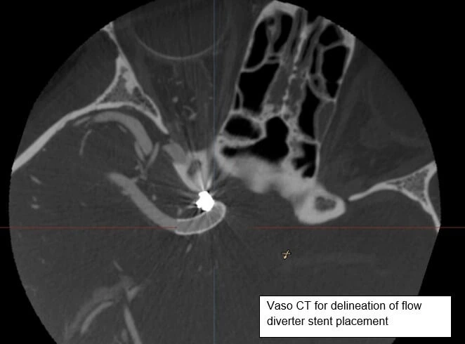
Vaso-CT ( of Philips Medical system) is a more higher resolution version of CBCT. During a Vaso-CT biplane angiography, a contrast agent is injected into the patient's bloodstream to highlight the blood vessels.
The additional advantage of Vaso-CT is it provides detailed information of the various devices placed inside the brain blood vessels including stents, flow diverter devices, intrasccular devices like Web.
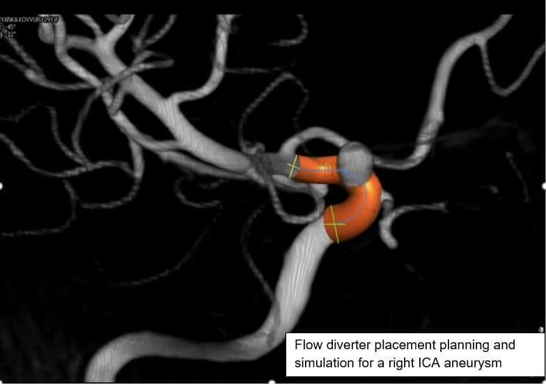
Specific details pertaining to device functioning like adequacy of device placement, impingement on normal brain blood vessels and adequate opening of the device are exclusively delineated using this particular software.
DR.SURESH GIRAGANI CONSULTANT INTERVENTIONAL RADIOLOGIST at Apollo Hospital, has more than Seventeen years of clinical experience in vascular interventions with a special interest in neurovascular and peripheral vascular disease interventional procedures.
Copyright 2021 Neuro All rights reserved. | Powered By KBK Business Solutions