
Image-guided thermal ablation that destroys liver, lung, kidney, and bone tumors with heat, delivered through a thin probe — often curative for small, early-stage disease.
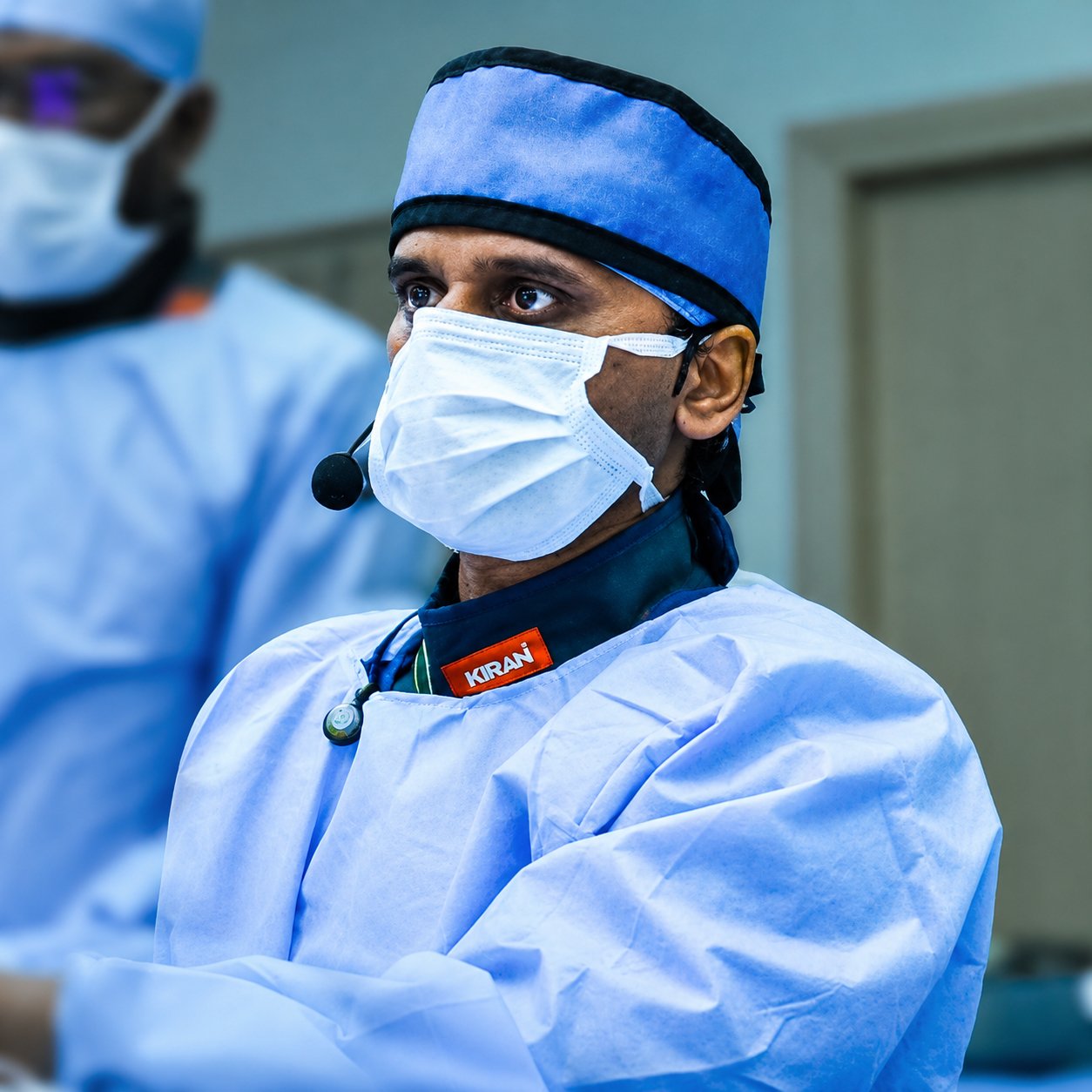
Both are thermal ablation techniques that destroy tumour tissue with heat, delivered through a thin probe or antenna inserted directly into the tumour under CT or ultrasound guidance. Radiofrequency ablation (RFA) passes an alternating electrical current through the tissue, generating heat through resistance as ions in the tissue vibrate around the probe tip. Microwave ablation (MWA) instead uses electromagnetic energy to agitate water molecules directly, generating heat more quickly and over a larger volume, and without needing to complete an electrical circuit through the body the way RFA does.
In practice, this means MWA tends to reach higher temperatures faster, creates a larger and more predictably shaped ablation zone, and is less affected by nearby blood vessels carrying away heat (the "heat-sink effect") than RFA — which is why microwave ablation has increasingly become the preferred technique, particularly for larger tumours or those near larger vessels, in centres where both are available. Both techniques share the same basic principle and the same core benefit: destroying a tumour precisely, from the inside, through a needle-sized access point rather than an operation.
Small hepatocellular carcinoma or selected metastases, potentially curative for early-stage disease under about 3cm.
Early-stage lung cancer in patients not fit for surgery, or a limited number of metastases.
Small renal masses under 3–4cm, a nephron-sparing alternative to surgery.
Painful bone metastases, or curative treatment of small benign tumours like osteoid osteoma.
Imaging maps the safest path to the tumour, avoiding vessels, nerves, and organs.
A thin probe is advanced directly into the tumour under real-time imaging guidance.
Heat is delivered for several minutes, destroying the tumour and a margin around it.
Most patients go home the same day or after one night.
Ablation is a well-established, guideline-supported treatment for small hepatocellular carcinoma (HCC) and selected liver metastases, particularly in patients who aren't candidates for surgical resection, and is considered potentially curative for very early and early-stage tumours generally under about 3 cm. A probe is placed directly into the tumour under CT or ultrasound guidance, and the surrounding tissue — including a margin of apparently healthy liver tissue around the tumour, to reduce the chance of microscopic disease being left behind — is heated to destructive temperatures. Most patients go home the same day or after a short overnight stay, and studies comparing the two techniques in the liver have generally found microwave ablation achieves a lower rate of local tumour regrowth than radiofrequency ablation, though both remain effective, well-established options.
In the lung, ablation is used primarily for patients with early-stage non-small cell lung cancer who aren't fit enough for surgery, or for a limited number of metastases from cancers elsewhere in the body, when surgery or radiotherapy isn't the right option for that individual. A probe is advanced through the chest wall directly into the tumour under CT guidance, with the patient under sedation or general anaesthesia. Because the probe passes through lung tissue to reach the target, the most common procedure-related risk is a pneumothorax — air leaking into the space around the lung — which is usually managed simply, sometimes with a small temporary chest tube, and doesn't typically prolong the hospital stay significantly. Ablation in the lung is generally reserved for smaller tumours and used as part of a broader treatment plan agreed with the patient's oncology and pulmonology team.
For small kidney tumours — typically renal cell carcinoma under about 3 to 4 cm — ablation offers a nephron-sparing alternative to surgery, particularly valuable for patients with reduced kidney function, tumours in both kidneys, multiple medical problems that make surgery riskier, or a strong personal preference to avoid an operation. A probe is guided into the tumour under CT or ultrasound guidance, and the kidney's naturally rich blood supply is factored into planning, since blood flow can carry heat away from the treatment zone. Kidney function is generally very well preserved after ablation compared with more extensive surgery, since only the tumour and a small margin of surrounding tissue are treated rather than removing a larger portion of the kidney.
Bone is a somewhat different application of the same technology, used in two distinct situations. The first is treatment of certain small, benign but persistently painful bone tumours, such as osteoid osteoma, where ablation is now considered a first-line treatment, often curing the pain in a single outpatient session. The second, and more common, use is palliative treatment of painful metastatic deposits in bone from cancers elsewhere in the body — here, the goal isn't necessarily to eliminate the cancer, but specifically to relieve pain that hasn't responded adequately to radiotherapy or medication, by destroying the pain-generating nerve fibres and tumour tissue within the bone. This can be genuinely transformative for a patient's quality of life, often significantly reducing pain within days, and is sometimes combined with cement injection into the treated bone (a technique similar to vertebroplasty) to add structural support where a bone is also at risk of fracture.
Planning: a CT, MRI, or ultrasound is reviewed beforehand to plan the safest, most direct path to the tumour, avoiding major blood vessels, nerves, and adjacent organs.
Access: under sedation or general anaesthesia, a thin probe or antenna is advanced directly into the tumour through the skin, guided in real time by CT, ultrasound, or a combination of both.
Ablation: heat is delivered for a set period — typically several minutes to around fifteen minutes per position, depending on tumour size — destroying the targeted tissue and a margin around it. Larger tumours may need more than one probe position, or an overlapping series of ablations, to cover the whole area.
Confirmation and recovery: imaging at the end of the procedure helps confirm the ablation zone covers the tumour, and patients are observed for a period afterward before going home, typically the same day or after one night in hospital.
Follow-up imaging — usually a contrast CT or MRI at around one month, then at intervals afterward — is used to confirm the treated area shows no residual or recurrent viable tumour, since an ablation zone continues to change in appearance as it heals, and distinguishing expected post-treatment change from true recurrence requires specific expertise. Regular surveillance imaging afterward is a standard part of ablation follow-up across all four organ systems, not a sign that anything has gone wrong.
Getting the ablation zone to fully cover the tumour with an adequate margin — while avoiding nearby vessels, nerves, airways, or bowel — depends on careful pre-procedure planning and precise, image-guided probe placement, and the right modality and approach differs meaningfully by organ and by tumour location. Decisions about which patients are better served by ablation versus surgery, radiotherapy, or systemic treatment are made jointly with oncology, surgery, and the relevant organ-specialist team, since ablation is one part of a broader cancer care pathway rather than a standalone decision.
Both destroy tumour tissue with heat delivered through a needle, but microwave ablation typically heats faster, creates a larger and more predictable ablation zone, and is less affected by nearby blood vessels carrying heat away — which is why it's increasingly preferred where both are available, though radiofrequency ablation remains a well-established, effective option too.
It depends on the situation. For small, early-stage liver, lung, or kidney tumours, ablation can be genuinely curative. For painful bone metastases from advanced cancer elsewhere in the body, the goal is usually pain relief and local control rather than a cure of the underlying disease.
Risks vary by organ — in the lung, the main risk is a pneumothorax (a small air leak around the lung), usually managed simply. In the liver and kidney, risks include bleeding or injury to a nearby structure. Overall, ablation carries a lower complication rate than open surgery for comparable tumours.
Because ablation targets only the tumour and a small margin around it, rather than removing a larger portion of the kidney as some surgical approaches do, kidney function is generally very well preserved afterward — this is one of the main reasons ablation is favoured for patients with reduced kidney function or tumours in both kidneys.
Many patients notice meaningful pain relief within days of the procedure, which can be a significant improvement in quality of life, particularly when pain hasn't responded well to radiotherapy or medication alone.
Book a consultation with Dr. Giragani, or send your scans for a specialist second opinion.