
What Happens During a DSA (Digital Subtraction Angiography) Procedure?
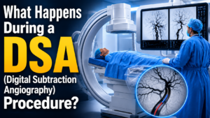
If you have been advised to undergo a DSA procedure in Hyderabad, understanding the process can help reduce anxiety and prepare you for the examination. Digital Subtraction Angiography (DSA) is considered the gold standard for diagnosing vascular disorders, including brain aneurysms, stroke, arteriovenous malformations (AVMs), carotid artery disease, and other blood vessel abnormalities.
For patients seeking advanced brain aneurysm diagnosis in Hyderabad or stroke evaluation in Hyderabad, DSA provides highly detailed images that help specialists accurately diagnose and plan the most effective treatment.
What Is DSA?
Digital Subtraction Angiography (DSA) is a minimally invasive imaging technique that uses X-rays and contrast dye to create highly detailed images of blood vessels.
Unlike standard X-rays, DSA removes background structures such as bones and soft tissues, allowing doctors to see blood vessels with exceptional clarity. This makes it one of the most accurate tools for diagnosing vascular diseases affecting the brain, neck, abdomen, and other parts of the body.
Why Is DSA Performed?
DSA plays a vital role in modern neurovascular imaging in Hyderabad and is frequently used to diagnose:
- Brain aneurysms
- Stroke and blocked arteries
- Arteriovenous malformations (AVMs)
- Carotid artery disease
- Vascular malformations
- Brain hemorrhage
- Peripheral vascular disease
- Blood vessel narrowing
DSA is also commonly performed before procedures such as:
- Aneurysm Coiling
- Flow Diverter Stent Placement
- Stroke Thrombectomy
- AVM Embolization
- Carotid Artery Stenting
As one of the most accurate forms of cerebral angiography, DSA allows the best interventional neuroradiologist in hyderabad to visualize blood vessels with exceptional precision.
Before the Procedure
Before undergoing a DSA test, your doctor will review your medical history and perform certain investigations, including:
- Blood tests
- Kidney function tests
- Allergy assessment
- Medication review
Patients may be advised to:
- Fast for several hours before the procedure
- Inform the doctor about blood-thinning medications
- Report any allergy to contrast dye
- Stay hydrated before the test
An intravenous (IV) line will be inserted to administer fluids and medications if required.
What Happens During a DSA Procedure?
Step 1: Preparation
You will be taken to a specialized angiography suite and asked to lie comfortably on the procedure table.
The procedure site, usually the wrist or groin, will be cleaned and sterilized. A local anesthetic is administered to numb the area.
Most patients remain awake during the procedure, although mild sedation may be provided for comfort.
Step 2: Catheter Insertion
A small puncture is made in an artery, commonly through:
- Radial artery (wrist)
- Femoral artery (groin)
A thin flexible catheter is inserted into the artery and carefully guided to the blood vessels being examined.
Using real-time X-ray guidance, the Interventional Neuroradiologist navigates the catheter safely to the target area.
Step 3: Contrast Dye Injection
Once the catheter reaches the desired location, a contrast dye is injected.
The dye makes blood vessels visible on X-ray images and helps create detailed vascular maps.
During this stage, some patients may briefly experience:
- Warm sensation
- Mild flushing
- Metallic taste in the mouth
These sensations are completely normal and disappear within seconds.
Step 4: Image Acquisition
As the contrast dye flows through the blood vessels, a series of high-resolution X-ray images is captured.
The digital subtraction technology removes surrounding tissues and bones from the images, producing exceptionally clear views of the vascular anatomy.
Doctors evaluate these images to identify:
- Brain aneurysms
- Blood vessel narrowing
- Stroke-causing blockages
- Arteriovenous malformations
- Abnormal blood flow patterns
- Vascular malformations
Step 5: Completion of the Procedure
After imaging is completed, the catheter is removed carefully.
Pressure is applied to the puncture site or a vascular closure device may be used to prevent bleeding.
Most DSA procedures take approximately 30 to 60 minutes, depending on the complexity of the condition.
What Happens After DSA?
Following the procedure, patients are monitored in a recovery area for a few hours.
Medical staff monitor:
- Blood pressure
- Heart rate
- Neurological status
- Puncture site
Patients are generally advised to:
- Drink plenty of fluids
- Avoid strenuous activities for 24–48 hours
- Keep the puncture site clean
- Follow medication instructions
Most patients can return home on the same day.
Is DSA Painful?
DSA is generally well tolerated and not painful.
Patients may feel:
- A slight prick during local anesthesia
- Mild pressure during catheter insertion
- Temporary warmth during contrast injection
Serious discomfort is uncommon.
What Are the Risks of DSA?
DSA is considered a very safe procedure when performed by experienced specialists.
Potential risks include:
- Minor bruising
- Bleeding at the puncture site
- Contrast dye allergy
- Infection
- Blood vessel injury
- Rare neurological complications
The overall complication rate remains very low.
Benefits of DSA
DSA offers numerous advantages:
- Gold standard vascular imaging
- Exceptional image quality
- Accurate diagnosis
- Real-time blood flow assessment
- Treatment planning assistance
- Ability to combine diagnosis and intervention
For patients undergoing brain aneurysm treatment in Hyderabad, DSA provides unmatched diagnostic accuracy and treatment guidance.
DSA for Brain Aneurysm Treatment in Hyderabad
Patients undergoing aneurysm coiling in Hyderabad or flow diverter treatment in Hyderabad often require DSA before and after treatment.
DSA helps specialists:
- Assess aneurysm size and shape
- Evaluate aneurysm neck anatomy
- Plan endovascular treatment
- Confirm successful aneurysm occlusion
- Detect recurrence during follow-up
As the gold standard for brain aneurysm diagnosis in Hyderabad, DSA remains an essential component of modern interventional neuroradiology. Dr. Suresh Giragani is one of the leading specialists in interventional neuroradiology in Hyderabad, with expertise in:
- Digital Subtraction Angiography (DSA)
- Aneurysm Coiling
- Flow Diverter Stent Placement
- Stroke Thrombectomy
- AVM Embolization
- Carotid Artery Stenting
- Neurovascular Interventions
With over 17 years of experience and more than 1,400 intracranial neurointerventions, he provides advanced diagnosis and treatment for complex neurovascular disorders.
Frequently Asked Questions (FAQs)
How long does a DSA procedure take?
Most DSA procedures take between 30 minutes and 1 hour, depending on the complexity of the condition.
Will I be awake during DSA?
Yes. Most patients remain awake, although mild sedation may be administered if necessary.
Is DSA better than CT Angiography or MR Angiography?
DSA is considered the gold standard because it provides the highest resolution images and real-time blood flow assessment.
Can I go home after DSA?
Most patients are discharged on the same day after a short observation period.
Is DSA safe?
Yes. DSA is generally very safe when performed by experienced Interventional Neuroradiologists.
Why is DSA important before aneurysm coiling?
DSA provides detailed information about aneurysm anatomy, helping specialists plan the safest and most effective treatment strategy.
Conclusion
Whether you are being evaluated for a brain aneurysm, stroke, or another vascular condition, Digital Subtraction Angiography (DSA) provides highly accurate information that guides diagnosis and treatment.
For patients seeking advanced
the procedure remains an essential component of modern neurovascular care, helping specialists deliver safe, effective, and personalized treatment.
About the Author:

Name: DR . SURESH GIRAGANI
INTERVENTIONAL RADIOLOGIST
DR. SURESH GIRAGANI CONSULTANT INTERVENTIONAL RADIOLOGIST at Apollo hospitals Jubilee Hills has more than Seventeen years of clinical experience in vascular interventions with a special interest in neurovascular and peripheral vascular disease interventional procedures.